Protein–protein & protein–peptide docking on Vecura
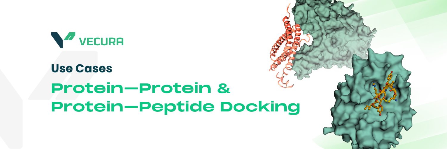
Vecura offers four operations for modelling how a protein binds another protein or a peptide, from fast geometric pose prediction to physics-guided integrative docking, ΔΔG mutation scanning, and restraint-file generation. The four operations are: protein-protein docking, protein-peptide docking, protein-protein binding change prediction, and docking restraint sampling. Choosing the right operation before selecting a model prevents fitting the wrong tool to the question. The sections below describe what each operation produces and which models serve it.
Choosing an operation and model
| What you have | What you want | Use | Typical runtime |
| Two folded protein structures | A ranked set of complex poses | protein-protein docking | Seconds (learning-based) to hours (integrative) |
| A protein structure + a peptide | A bound peptide pose in a defined pocket | protein-peptide docking | Minutes |
| A protein–protein complex PDB + mutation list (or WT + mutant complex PDBs) | ΔΔG (kcal/mol) per mutation | protein-protein binding change prediction | Seconds–minutes |
| Two chains' active/passive residue lists | A CNS-format restraint file (.tbl) | docking restraint sampling | Seconds |
The biggest practical trade-off is speed vs. accuracy and flexibility. Rigid-body deep-learning models (EquiDock, AF2Dock) are the fastest and need no MSA, but they keep backbones rigid and accept no experimental restraints. Not every learning-based model shares those limits: GeoDock allows flexibility, and ColabDock accepts restraints. Physics-aware integrative methods (HADDOCK3) are slower but combine experimental restraints with flexible interface refinement and return ranked, refined ensembles rather than a single pose.
The operations
Protein-protein docking
Protein-protein docking
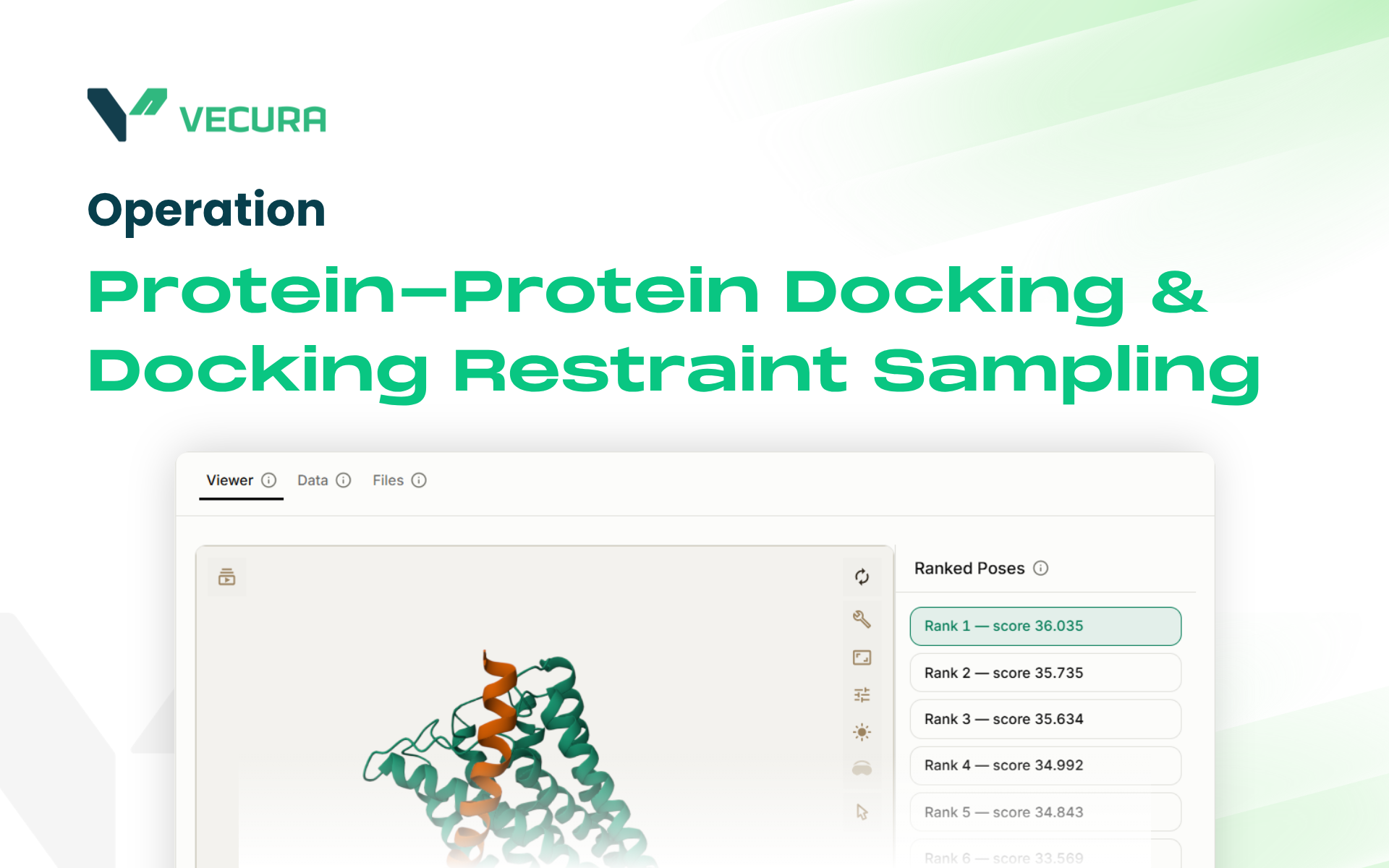 Use case replayProtein-Protein Docking & Docking Restraint SamplingSee this use case
Predicts the 3D complex structure formed when two folded protein chains bind.
Use this operation, not protein-peptide docking, when both partners are folded domains; the platform does not block peptide inputs here, but these models are trained and benchmarked on folded proteins, so peptide results are less reliable. Choose EquiDock or AF2Dock for speed, GeoDock when flexibility matters, ColabDock or LightDock when you have experimental restraint data, and HADDOCK3 when you need rigid-body sampling followed by flexible refinement in one pipeline.
Input
Two protein structures (PDB)
Output
Ranked docked complex(es) (PDB)
Which model
Six models cover this operation, and choosing between them is really choosing where you sit on the tradeoff above: EquiDock or AF2Dock for speed, GeoDock when flexibility matters, ColabDock or LightDock when you have experimental restraint data, and HADDOCK3 when you need rigid-body sampling followed by flexible refinement in one pipeline.
| Model | Approach | Reach for it when | ||
| LightDock | GSO swarm-based docking; accepts residue-level restraints and multiple scoring functions; also handles protein-peptide and protein-DNA in its underlying framework | You want a swarm sampler with explicit restraint support | ||
| EquiDock | SE(3)-equivariant rigid-body docking in a single forward pass; no MSA, no restraints | You need the fastest geometry-only prediction | ||
| ColabDock | AlphaFold2-Multimer with 1v1, 1vN, MvN, and repulsive restraint types integrated into the prediction | You have experimental data (crosslinks, NMR CSPs, mutagenesis) to guide the run | ||
| HADDOCK3 | Modular integrative platform (BonvinLab, Utrecht): rigid-body sampling, semi-flexible refinement, scoring; also generates restraint files (see docking restraint sampling, below) | You want physics-aware integrative docking with full experimental-restraint support | ||
| GeoDock | Residue-level flexible docking; no MSA required; can score a predicted complex against a native reference using DockQ | You are docking unbound (apo) structures and need flexibility during docking, without an MSA | ||
| AF2Dock | AlphaFold-Multimer backbone adapted for flow-matching docking; MSA-free inference | You want an AF2-Multimer-style backbone without the MSA overhead |
Protein-peptide docking
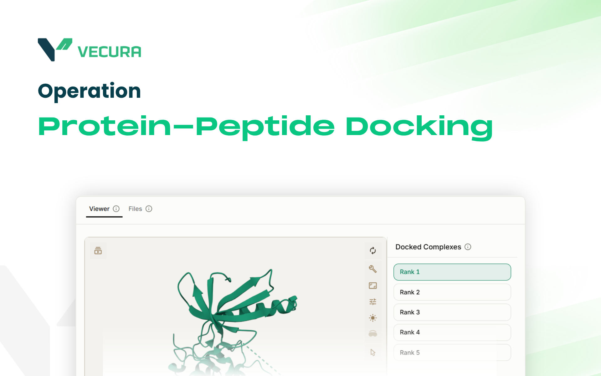 Use case replayProtein-peptide dockingSee this use case
Protein-peptide docking places a short peptide into a defined pocket on a protein receptor using a generative diffusion process.
Use this instead of protein-protein docking when one partner is a peptide (input as PDB) and the binding site can be defined by a reference ligand or pocket-centre coordinates. The pocket radius (5–25 Å) and number of poses (1–200) are configurable.
Input
Receptor protein PDB + peptide PDB(s); pocket defined by reference ligand (SDF/MOL2) or XYZ centre
Output
Docked peptide pose(s) (PDB), up to 200 per peptide
Which model
PocketXMol (Docking) is the only model here: a pocket-interacting generative diffusion model that docks peptides into a protein pocket defined by a reference ligand or an explicit XYZ centre, generating up to 200 poses. Reach for it when you have a pocket-defined receptor and a peptide structure to place.
Protein-protein binding change prediction
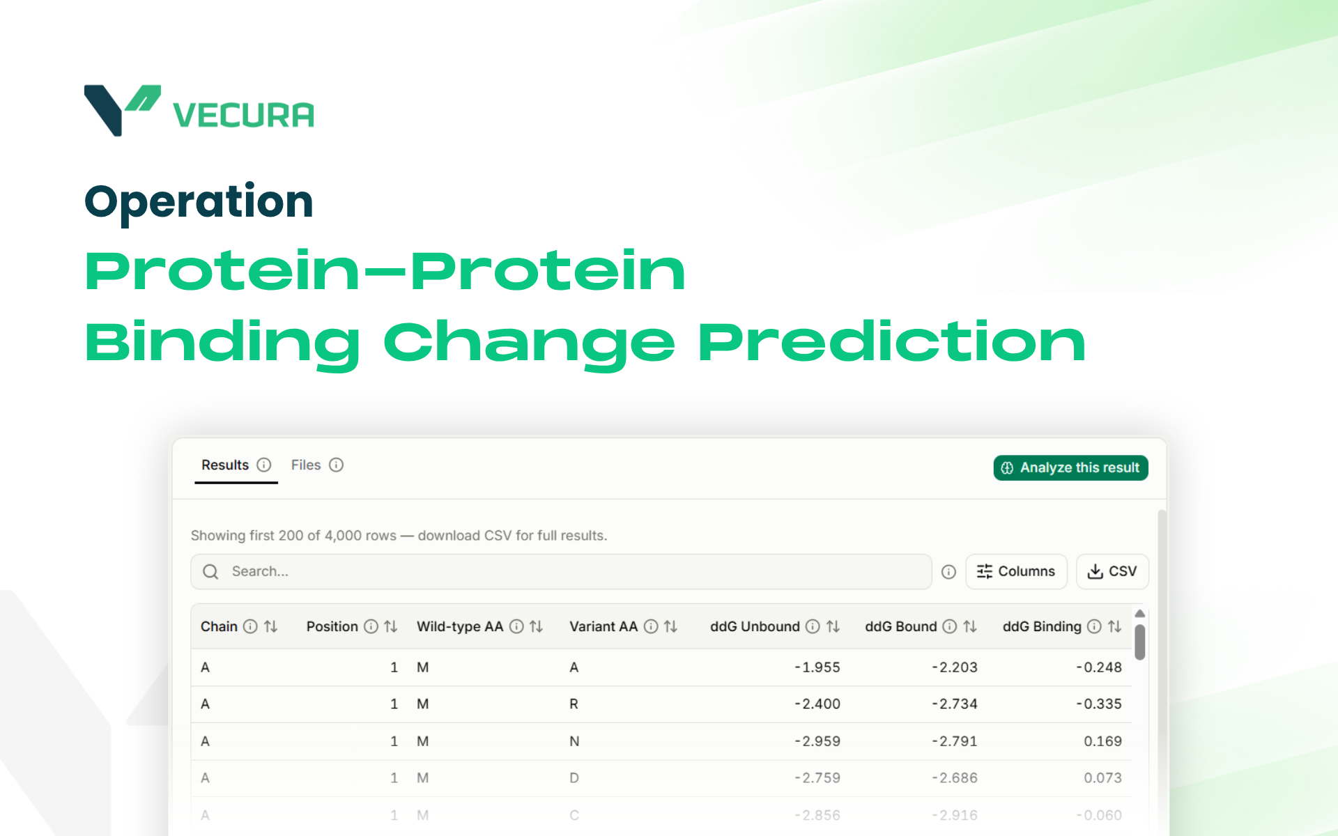 Use case replayProtein-Protein Binding Change PredictionSee this use case
Protein-protein binding change prediction estimates the change in binding free energy (ΔΔG, kcal/mol) caused by one or more point mutations.
Use it to rank interface mutations, stabilising vs. destabilising, as a rapid in silico screen before experimental mutagenesis. Neither model replaces biophysical measurement; treat outputs as a prioritisation filter. Check each model's sign convention (whether a positive ΔΔG means weaker or stronger binding) before ranking mutations.
Input
(StaB-ddG): Complex PDB + mutation strings (format: , e.g. EA63Q) (DDG Predictor): Wild-type complex PDB + mutant complex PDB (pre-built, same residue count)
Output
ΔΔG (kcal/mol) per mutation
Which model
The two models differ mainly in what they need from you.
-
StaB-ddG is a ProteinMPNN-based ΔΔG predictor that works from a single complex PDB plus a mutation list, with three checkpoints: dual fine-tuned (recommended), stability-only, and base; take it when you want fast ΔΔG scanning across many point mutations from one structure.
-
DDG Predictor is a geometric-attention deep-learning model that requires a pre-built wild-type PDB and a pre-built mutant PDB (e.g. from Rosetta) and returns a single ΔΔG value; take it when you already have both complex structures in hand.
Docking restraint sampling
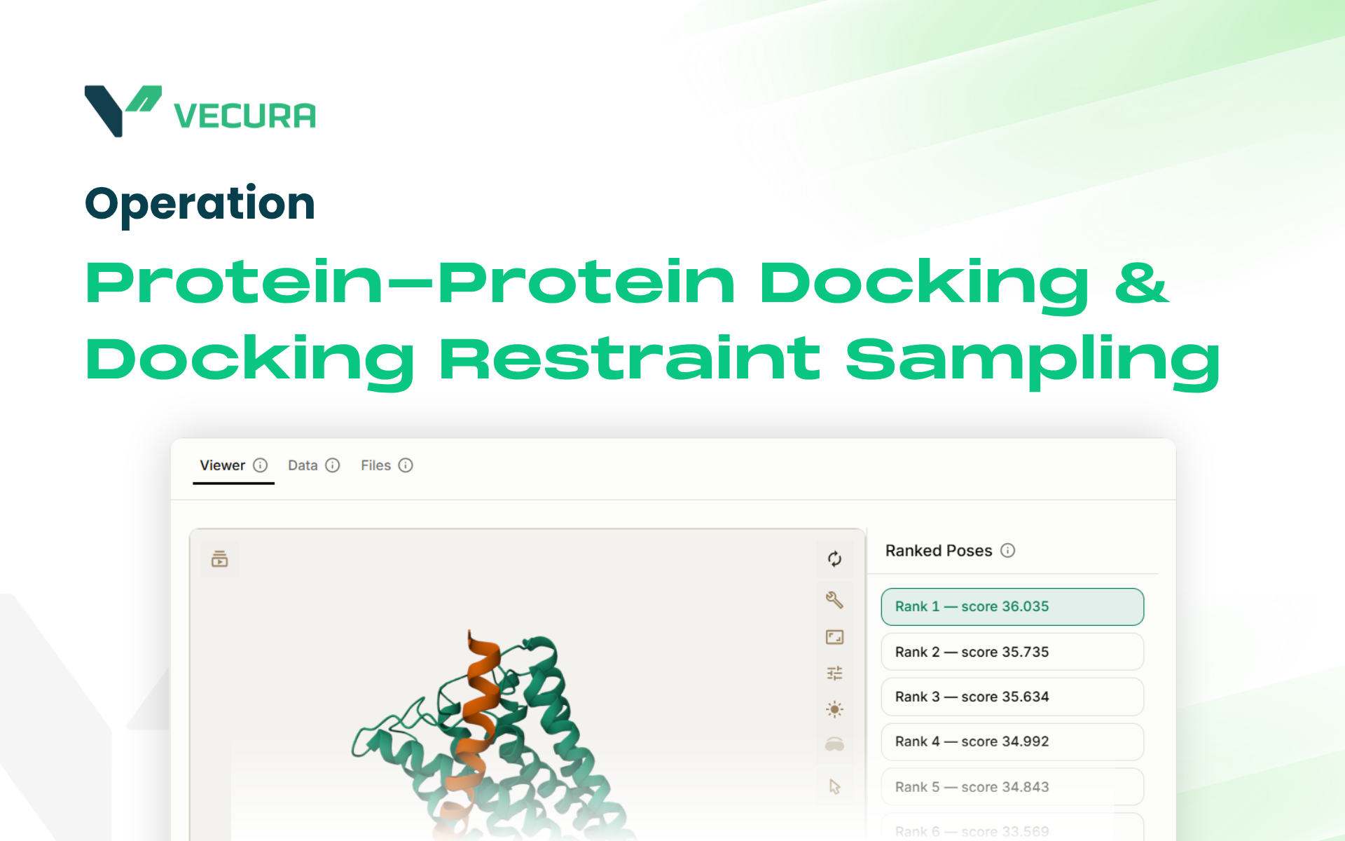 Use case replayProtein-Protein Docking & Docking Restraint SamplingSee this use case
Docking restraint sampling converts residue-level interface information, active residues (confidently at the interface) and passive residues (nearby/probable) for each chain, into a CNS-format ambiguous interaction restraints file (.tbl).
This is a preparatory step, not a docking method: no complex structure is produced, and the output feeds into a subsequent HADDOCK3 protein-protein docking run.
Input
Chain IDs and active/passive residue lists for two binding partners
Output
CNS-format .tbl restraint file
Which model
HADDOCK3 is the only model here, the same integrative platform listed under protein-protein docking, used in its restraint-generation mode. It turns active and passive residue lists for two chains into a .tbl file that can feed directly into its own docking run.
Next step
After obtaining a docked complex, a natural downstream step is to score the interface with a binding-affinity predictor (PRODIGY for protein–protein affinity) or to scan interface mutations with protein-protein binding change prediction. For lead optimization, MD simulation of the bound complex tests whether the pose is stable; ADMET property prediction applies mainly when the binder is a peptide or small molecule.