How to Slow down Amyotrophic Lateral Sclerosis - An In-Silico Approach to SOD1 Stabilization
A proposed computational pipeline for identifying small-molecule binders at the SOD1 dimer interface
How to Slow down Amyotrophic Lateral Sclerosis - An In-Silico Approach to SOD1 Stabilization
A proposed computational pipeline for identifying small-molecule binders at the SOD1 dimer interface
The Problem: ALS and the Unmet Need
Amyotrophic lateral sclerosis (ALS) is a devastating neurodegenerative disease. It kills motor neurons - the cells that control voluntary movement - leading to progressive paralysis. Most patients die within 3-5 years of diagnosis. There is no cure, and the few approved treatments offer only modest benefits.

Figure 1. ALS and the loss of motor-neuron function. Healthy motor neurons (left) innervate muscle and drive contraction; in ALS, motor-neuron degeneration (right) interrupts that signal, leaving the muscle unable to contract and producing progressive paralysis (PC: York Rehab Clinic)
Riluzole, approved in 1995, extends survival by approximately 2-3 months. Edaravone, approved in 2017, slows functional decline in a subset of patients. Neither stops the disease. The need for fundamentally new therapeutic approaches is urgent.
What SOD1 does normally
SOD1 is a 153-residue metalloenzyme that converts toxic superoxide radicals into oxygen and hydrogen peroxide - a critical antioxidant defense. It functions as a homodimer, with each subunit binding one copper ion and one zinc ion. The copper drives the catalytic reaction; the zinc stabilizes the protein's structure. A disulfide bond (Cys57-Cys146) further locks each subunit into its native fold.

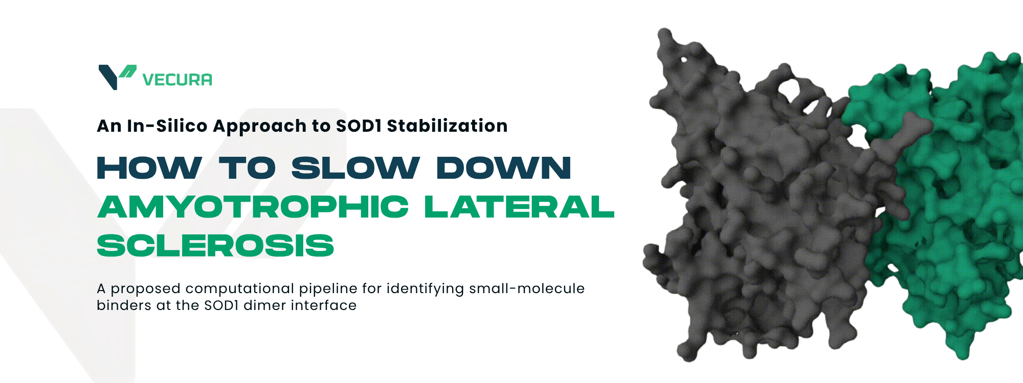
Figure 2. The native SOD1 homodimer. Molecular-surface view of the holo (Cu/Zn-bound) human SOD1 dimer, with the two identical subunits shaded separately to show the extensive subunit-subunit interface that the screen aims to stabilize (PDB 1HL5).
The aggregation pathway: a drug target
The aggregation cascade is the most druggable aspect of SOD1 pathology.
It follows a recognizable sequence:

Figure 3. The SOD1 aggregation cascade and its intervention points. Destabilization of the native dimer releases monomers that misfold, self-associate into toxic soluble oligomers, and mature into insoluble amyloid fibrils.
Each step in this cascade is a potential intervention point. But the most attractive one for small-molecule drug design is step 1: stabilizing the dimer before the cascade even begins.
The Strategy: Structure-Based Virtual Screening

Figure 4. End-to-end in-silico screening workflow
The idea is straightforward: take a high-resolution 3D structure of SOD1, identify pockets where a drug could bind, dock thousands of small molecules into those pockets, and validate the best hits with molecular dynamics. Every step is computational: no lab bench required for the initial screening. You can try the workflow on Vecura Platform here.
Here's the pipeline at a glance:
-
Structure preparation: Get the best SOD1 structure, clean it up
-
Pocket identification: Find where a drug could bind
-
Compound library: Assemble a focused set of drug-like molecules
-
Molecular docking: Score each compound for binding
-
Hit prioritization: Filter, cluster, and rank the best binders
-
Molecular dynamics: Binding validation
Let's walk through each step.
Step 1: Structure Preparation
The first decision is which structural state of SOD1 to target. This is not merely a technical consideration - it is a strategic choice that reflects the underlying therapeutic hypothesis. The two states answer different biological questions:

Figure 5. Choosing which structural state to target.
Both strategies are biologically valid targets - pick the one matching your intended clinical stage, not whichever screens better. For the present workflow, the holo SOD1 structure (PDB: 2C9V) was selected. The objective is to identify compounds capable of stabilizing the native Cu/Zn-bound dimer and reducing its propensity to dissociate into aggregation-prone species.
The holo monomers in 2C9V (chains A and F) were both retained to preserve the biological dimer. Crystallographic waters and non-biological crystallization artifacts (sulfate and sodium ions) were removed, while the catalytic Cu and Zn ions were kept, as they are integral to the native protein structure. Alternate atom conformations were resolved by retaining only the highest-occupancy coordinates. Hydrogens were then added with protonation states assigned for physiological pH (7.4).

Figure 6. Receptor preparation of PDB 2C9V.
The resulting structure serves as a clean, biologically relevant receptor model for subsequent virtual screening and docking studies.
Step 2: Pocket Identification
Decades of SOD1 research already tell you which regions matter, so the choice is whether to define pockets from that prior knowledge or let a geometric algorithm find them blind.

Figure 7. Knowledge-driven versus algorithm-driven pocket identification.
For this work, the dimer interface was selected as the primary docking region. This choice is directly aligned with the therapeutic objective of stabilizing the native Cu/Zn-bound SOD1 dimer and preventing the initial dissociation event that precedes misfolding and aggregation.
Step 3: Compound Library
Vecurate, a focused library of over 700,000 natural products, was passed through a four-stage filtering pipeline. Compounds were first screened against Lipinski's Rule of Five and rejected if they incurred more than one violation (MW > 500, LogP > 5, HBD > 5, HBA > 10), keeping the library in drug-like chemical space. A PAINS filter then removed pan-assay interference compounds - structural motifs such as quinones, catechols, and rhodanines that react non-specifically and surface as false positives across many targets. Each surviving compound was assigned a low-energy 3D structure using RDKit's ETKDGv3 algorithm with MMFF94 force-field optimization.
Step 4: Molecular Docking
Docking was run with SigmaDock, a fragment-based SE(3) Riemannian diffusion model for molecular docking
| Parameter | Value | Rationale |
| Binding site center | (20.0, 0.0, 14.0) | Geometric midpoint of dimer interface (chains A and F) |
| Binding site radius | 5 Å | Defines pocket residue context for the diffusion model |
| Poses per compound | 5 | Top-5 poses sampled per ligand |
| Rescoring | Vinardo (GNINA) | Physics-based rescoring for final ranking |
| PoseBusters check | Enabled | Filters chemically implausible poses |
For each compound, the model reports a binding affinity in kcal/mol (more negative = stronger binding) and the 3D coordinates of the predicted binding pose.
Step 5: Hit Prioritization
Docked compounds were sorted into three affinity tiers:
| Tier | Affinity (kcal/mol) | Interpretation |
| Tier 1 | ≤ −8.0 | Strong binders |
| Tier 2 | −8.0 to −7.0 | Moderate binders |
| Tier 3 | −7.0 to −6.0 | Borderline binders |
Surviving hits were then clustered by structural similarity using Morgan (circular) fingerprints, which encode each atom's local environment, and the Butina algorithm with a Tanimoto distance cutoff of 0.3 (i.e. compounds ≥ 0.7 similar are grouped together) . From each cluster, the compound with the best affinity was taken as the representative, so the final list stays chemically diverse rather than ten near-identical variations on one scaffold.
Step 6: Molecular Dynamics - Does the Compound Actually Stay Bound?
Docking is an excellent starting point, but it's only a snapshot. It predicts how a ligand might fit into a protein at one instant in time. Real proteins, however, are anything but static. At body temperature (37°C), every atom is constantly vibrating, side chains flex, loops move, and ligands can shift or even drift away from their binding site.
This matters even more for SOD1 because we're targeting its dimer interface-a broad, shallow groove rather than a deep binding pocket. A ligand may receive an excellent docking score simply because it fits well in that frozen snapshot, yet once thermal motion begins, nothing prevents it from sliding off the surface. Docking alone can't distinguish between compounds that truly remain bound and those that only appear promising.
That's where molecular dynamics (MD) comes in. Instead of evaluating a single pose, MD simulates the behavior of every atom over time, allowing us to watch the protein and ligand interact under conditions that closely resemble the cellular environment. If a compound remains bound for hundreds of nanoseconds despite constant thermal fluctuations, we can be much more confident that the interaction is genuine.
Building a realistic simulation
Creating an MD simulation isn't as simple as pressing "Run." Every component of the system needs an accurate physical description.
First, the protein is prepared using the AMBER99SB-ILDN force field, which defines how standard amino acids behave during the simulation. The ligand is parameterized separately using GAFF2 with AM1-BCC charges so its atoms interact realistically with the protein and solvent.
The most technically challenging part is the Cu/Zn metal center. SOD1 relies on tightly coordinated copper and zinc ions to maintain its native structure. Standard force fields treat these ions as simple charged particles, which often causes them to drift away from their coordinating residues during a simulation. Once that happens, important structural elements-including the zinc-binding and electrostatic loops-can collapse for purely computational reasons rather than because the protein is genuinely unstable.
To avoid this artifact, the metal center is parameterized using MCPB.py (Metal Center Parameter Builder). This workflow derives bonded parameters from quantum mechanical calculations, preserving the native coordination geometry throughout the simulation.
Once every component has been parameterized, the protein, ligand, and metal center are combined into a single system, immersed in a box of water, and supplemented with sodium and chloride ions to reproduce physiological salt concentrations.
Running the simulation
Before the real simulation begins, the system undergoes a series of preparation steps.
Energy minimization removes steric clashes that may have been introduced while assembling the model. Short equilibration simulations then gradually bring the system to physiological temperature (310 K) and pressure (1 bar), allowing the solvent and ions to settle while the protein remains restrained.
Only after the system is fully equilibrated are all restraints removed for the production simulation.
For this workflow, production simulations typically run for 200–500 nanoseconds. Modern GPUs make this surprisingly practical-a 200 ns simulation of the SOD1 dimer, containing roughly 32,000 atoms including water, can usually be completed in 2–4 hours.
What are we looking for?
The trajectory generated by MD contains thousands of snapshots showing how the complex evolves over time. Several analyses help determine whether a compound is genuinely stable.
| Metric | Why it matters | Good sign |
| Ligand RMSD | Does the ligand stay in the same binding pose? | Stable plateau below ~2 Å |
| Protein–ligand contacts | Are key interactions maintained? | Contacts remain throughout the simulation |
| Hydrogen-bond occupancy | Do important hydrogen bonds persist? | High occupancy (>30%) |
| MM/PBSA binding energy | Is binding energetically favorable? | Consistently negative values |
No single metric tells the whole story. Instead, they build a consistent picture. A ligand that remains stable, preserves its key interactions, and maintains favorable binding energy is much more convincing than one that only achieved a good docking score.
Beyond the Dimer Interface
If the dimer interface screen doesn't yield validated hits (a possible and scientifically informative outcome), the pipeline can be redirected:
-
Screen the electrostatic loop: this region (residues 121-142) is more flexible and may form a more druggable pocket when destabilized
-
Screen against the apo form: PDB 1HL4 captures the aggregation-prone state; compounds that stabilize this conformation could prevent oligomerization.
-
Consider covalent inhibitors: SOD1's surface-accessible free cysteines offer a handle for covalent warheads.
-
Explore alternative modalities: antisense oligonucleotides (the Tofersen approach), protein-protein interaction stabilizers, or proteolysis-targeting chimeras (PROTACs)
A negative result at the dimer interface is not a failure, it's data that narrows the search space and redirects effort toward more promising strategies.
The Bigger Picture
This pipeline represents a practical, reproducible approach to computational drug discovery that can be applied to any protein target with a known structure. The key principles:
-
Start with the best structural data (high-resolution, physiologically relevant state)
-
Match the structural state to your therapeutic hypothesis (holo for dimer stabilization, apo for aggregation interception)
-
Use focused libraries (natural products)
-
Apply rigorous filtering (Lipinski, PAINS, chemical diversity)
-
Treat negative results as informative (they tell you where NOT to look)
For SOD1 specifically, this approach could identify new chemical starting points for ALS drug development or definitively show that the dimer interface is not the right target, saving years of wasted effort.
Reference
-
Rosen DR et al. Mutations in Cu/Zn superoxide dismutase gene are associated with familial amyotrophic lateral sclerosis. Nature. 1993;362(6415):59-62.
-
Kaur S, McKeown S, Rashid S. Mutant SOD1 mediated pathogenesis of Amyotrophic Lateral Sclerosis. Gene. 2016;577(2):109-118.
-
Saccon RA, Bunton-Stasyshyn RK, Fisher EM, Fratta P. Is SOD1 loss of function involved in amyotrophic lateral sclerosis? Brain. 2013;136(Pt 8):2342-2358.
-
Anzai I et al. A misfolded dimer of Cu/Zn-superoxide dismutase leading to pathological oligomerization in amyotrophic lateral sclerosis. Protein Science. 2017;26(3):484-496.
-
Wang LQ et al. Amyloid fibril structures and ferroptosis activation induced by ALS-causing SOD1 mutations. Science Advances. 2024;10(46):eado8499.
-
Tafuri F et al. SOD1 misplacing and mitochondrial dysfunction in amyotrophic lateral sclerosis pathogenesis. Frontiers in Cellular Neuroscience. 2015;9:336.
-
Peggion C et al. SOD1 in ALS: Taking Stock in Pathogenic Mechanisms and the Role of Glial and Muscle Cells. Antioxidants. 2022;11(4):614.
-
Blair HA. Tofersen: First Approval. Drugs. 2023;83(11):1039-1045.
-
Miller TM et al. Trial of Antisense Oligonucleotide Tofersen for SOD1 ALS. New England Journal of Medicine. 2022;387(12):1099-1105.
-
Miller TM et al. Phase 1-2 Trial of Antisense Oligonucleotide Tofersen for SOD1 ALS. New England Journal of Medicine. 2020;383(2):109-119.
-
Hamad AA et al. Tofersen for SOD1 amyotrophic lateral sclerosis: a systematic review and meta-analysis. Neurological Sciences. 2025;46(3):7994.
-
Benatar M et al. Design of a Randomized, Placebo-Controlled, Phase 3 Trial of Tofersen Initiated in Clinically Presymptomatic SOD1 Variant Carriers: the ATLAS Study. Neurotherapeutics. 2022;19(4):1248-1258.
-
Capper MJ et al. The cysteine-reactive small molecule ebselen facilitates effective SOD1 maturation. Nature Communications. 2018;9(1):1693.
-
Watanabe S et al. Ebselen analogues delay disease onset and its course in fALS by on-target SOD-1 engagement. Scientific Reports. 2024;14:16503.
-
Ip P et al. Quercitrin and quercetin-3-beta-D-glucoside as chemical chaperones for the A4V SOD1 ALS-causing mutant. Protein Engineering Design and Selection. 2017;30(7):477-484.
-
Hossain MA et al. Evaluating protein cross-linking as a therapeutic strategy to stabilize SOD1 variants in a mouse model of familial ALS. PLOS Biology. 2024;22(5):e3002462.
-
Wright GSA et al. Ligand binding and aggregation of pathogenic SOD1. Nature Communications. 2013;4:1758.
-
Li P, Merz KM Jr. MCPB.py original paper (JCIM, 2016)
-
Li P, Merz KM Jr. MCPB.py tutorial with GROMACS support (Methods Mol. Biol., 2021)
-
Prat A, Zhang L, Deane CM, Teh YW, Morris GM. SigmaDock: Untwisting Molecular Docking with Fragment-Based SE(3) Diffusion. ICLR 2026. arXiv:2511.04854. doi:10.48550/arxiv.2511.04854
-
Quiroga R, Villarreal MA. Vinardo: A Scoring Function Based on Autodock Vina Improves Scoring, Docking, and Virtual Screening. PLOS One. 2016;11(5):e0155183. doi:10.1371/journal.pone.0155183
Try Vecura now.
Bring your own inputs and start exploring what Vecura can do.